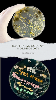
“Bacterial colony morphology is the fundamental step for characterization and identification of bacteria“
In order to study microbes, we grow the bacteria in laboratory. We cultivate the bacteria in solid and liquid media. In solid media, the bacteria grow in the form of colonies. Colony is a dot-like structure that appears on the surface of the solid media. The colony is formed due to the multiple multiplications of single bacteria (mother cell). The mother cell undergoes binary fission and produces identical daughter cells forming a colony. Hence, the colony consists of genetical clones of the mother cell.
The different bacterial species produces different shapes, surfaces, color, texture and size of the colony. Hence, this colony characteristic is used to identifying and classification of bacterial species. The study of bacteria colony is called colony morphology. The type of media and environmental factors used for bacterial cultivation affects the colony morphology. When an unknown bacterium is isolated, colony morphology is the first parameter used for its characterization.
The colony may have different shape, color, and sizes and hence colony morphology can be used for classification and identification of the bacteria. Following parameters are used to study bacteria colony –
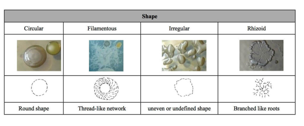
- Form or shape – circular, filamentous, irregular or rhizoid
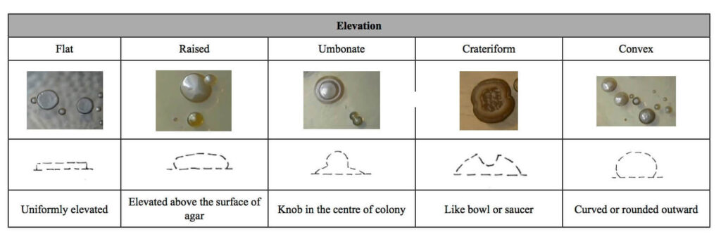
- Elevation (side view of colony) –
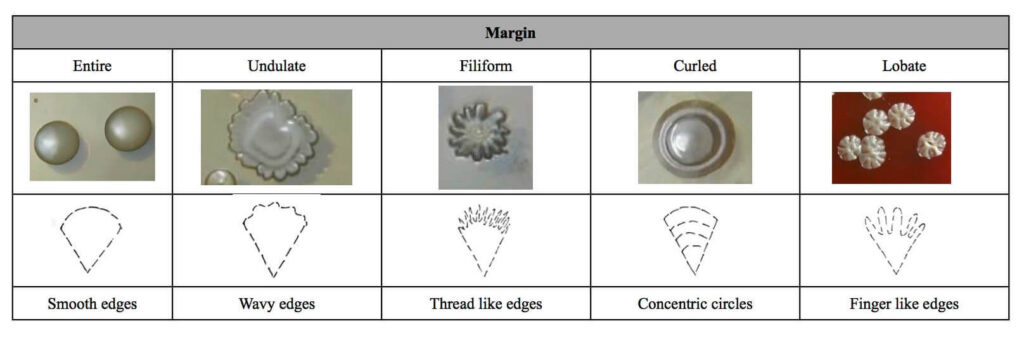
- Margin (border or edge of the colony)
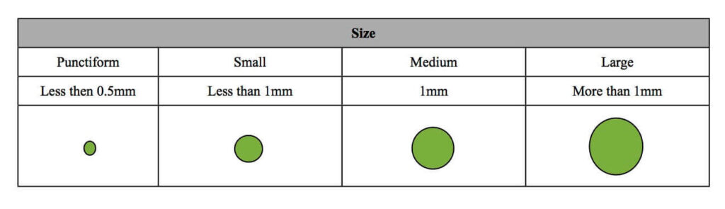
- Size – diameter varies and usually measured in milimeters.
- Opacity
- Surface
- Color
Colony Morphology: Shape
Elevation –
Colony Size –
The bacterial colony size varies from bacterial species to species. The size of the colony can be determined by measuring its diameter. The bacterial colony sizes are in the scale of milimeter. Based on the diameter size, the colony could be punctiform (less than 0.5mm), small (less than 1mm), medium (1mm) and large (more than 1mm).
Opacity –
The bacterial colony also varies in optical properties. Based on the opacity, the bacterial colonies are of following types –
- Opaque – Non transparent (cannot see through it)
- Translucent – Semi transparent (partially can be seen)
- Transparent – Can clearly see through it
- Iridescent – Shows different color in light reflection
Texture –
The bacterial colony surface texture appears different from bacterial species to species. The texture can be studied by touching the inoculating loop over the colony surface. Based on the observation, following texture has been noticed –
- Mucoid (mucus like)
- Slimy and moist (semi –liquid)
- Viscous and shiny
- Dry
Colony Color –
The bacterial colony are found in almost all colors. The color of all colony is due to the pigments released by the bacteria. The colony color is affected by the type of medium and nutrients provided.
Protocol to study the Colony Morphology –
The bacteria are omnipresent. They are present on our body surface, inside our body, air, soil, water etc. The bacteria can be isolated from any of the mentioned source. Suppose, you are isolating the bacteria from your air then prepare the nutrient media, autoclave it and pour it in sterile petri plate. Expose the nutrient media petri plate to air for few minutes and then close its lid. Incubate the plate in bacteriological incubator for 24 hours at 37°C. After incubation, count and label the colonies. Study the labeled colonies on aforementioned criteria and prepare your observation table.
After the studying the characteristics of the colonies, discuss it with your teacher and try to identify them.
Takeaways –
Studying bacteria colony morphology is the fundamental step for characterization and identification of the bacteria. It is also used for bacterial classification.
References –
- https://bio.libretexts.org/Learning_Objects/Laboratory_Experiments/Microbiology_Labs/Microbiology_Labs_I/08%3A_Bacterial_Colony_Morphology
- https://guides.baker.edu/c.php?g=303096&p=2022446
- https://www.cdc.gov/labtraining/docs/job_aids/biochemicals_gram_positive_organism_id/Colonial_Characteristics_Branded_508.pdf
- https://www.easynotecards.com/notecard_set/30494
- https://www.microbiologyinpictures.com/bacterial%20colonies.html
Dr. Sangha Bijekar has 7 years of Teaching Experience at University level. She loves to get engage in teaching and learning process. She is into blogging from last two years. She intends to provide student friendly reading material. She is avid Dog Lover and animal rescuer. She is learned Bharatnatyam and Katthak Dancer. She is into biking and She also loves to cook.