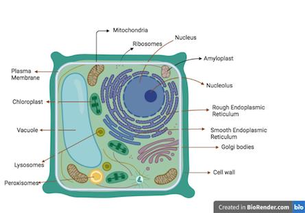
Introduction –
Plants are multicellular organisms and their cells are eukaryotic. Like animal cells, the plant cells have membrane bound organelles and a nucleus. However, the plant cell differs from animal cells in many aspects. The plant cells have some special organelles that allows the cells to carry out photosynthesis. Hence, the plant cell can be defined as a basic unit of plants, they are eukaryotic cell bordered by a cell wall and encloses membrane bound organelles that perform various functions.
Plant cell structure –
The plant cells are mostly found in rectangular shapes. Their size is larger than the animal cells. The plant cell and animal cells have similar organelles like cytoplasm, nucleus, ribosomes, mitochondria, golgi bodies, endoplasmic reticulum. However, the plant cells have special organelles like cell wall, vacuole and plastids (chloroplast, amyloplast and chromoplast). The organelles that are common in plant and animal cells have found to perform same functions in both types of cell.
Cell Wall –
- The plant cell is bordered and protected with the cell wall. The cell wall is absent in animal cells.
- The presence of a cell wall provides tensile strength that protects the cell from osmotic stress and mechanical damage.
- The cell wall is composed of network of cellulose microfibrils and the cross linking glycans are embedded in a highly cross linked matrix of pectin polysaccharides.
- The multiplying cells like meristem are small is size and have thin and extensible cell wall called primary cell wall. Once the cell is matured, the secondary cell wall is produced over primary one. And the secondary cell wall is rigid and non extensible. The chemical composition of primary and secondary cell wall is same however, the secondary cell wall is thick and contains lignin.
- During drought, the vacuoles may shrink but the rigid cell wall maintains the structural integrity of the plant cells.
- The plant cell wall is made up of cellulose fiber (polymer of glucose). The 40 cellulose fibers aggregate to form microfibrils. These microfibrils form a network with other polysaccharides and are cross linked to each other. Their strength is comparable to steel.
Functions of Cell wall –
- The primary function of cell wall is to mechanically protect the cellular content from the external environment.
- The cell wall is semi permeable in nature allowing exchange of water, ions, minerals and nutrients.
- The cell wall contains some regulatory molecules that plays a vital role in recognizing the pathogen.
- The fluid inside the plant cell produces a pressure called Turgor pressure that presses the plasma membrane against the cell wall.
- The turgor pressure is developed due to the presence of the cell wall and this provides rigid structure to the cell.
- When there is sufficient water, the vacuoles swell and produce more turgor pressure.
Cytoskeleton –
The network of filamentous proteins called cytoskeleton holds the cellular structure together. The cytoskeleton plays a vital role in maintaining the spatial organization of the cell, cell shape and during the cell division. There are three types of cytoskeleton proteins – microfilament, microtubule and intermediate filament.
Microtubules –
- The microtubules are made from the assembly of globular proteins.
- They are hollow cylinders with a diameter of 25nm.
- The microtubules are the polymer of tubulin protein.
- The tubulin (globular) protein is a heterodimer made up of α- and β-tubulin subunits.
- While forming a single microtubule, the thousands of tubulin repetitive monomers are arranged in 13 columns in the same direction forming protofilaments.
- Due to such arrangement, at one end there are only alpha subunits while on the other there will be beta subunits. Hence, their ends are polar.
- During cellular processes like mitosis, meiosis and cytokinesis, the length of microtubule filament needs to be increased or decreased called assembly and disassembly of microtubule respectively.
- Adding or removing tubulin monomers can achieve this. The end at which tubulin is added is called plus end while in which the tubulin monomers are removed, that end is called minus end.
Microfilament –
- The microfilaments are also made of an assembly of globular protein.
- Unlike Microtubules, they are solid (not hollow).
- Their diameter is 7nm, which is less than microtubule.
- The microfilaments are polymers of globular actin protein (G-actin).
- G-action is a single polypeptide chain.
- The microfilament is formed when two G-actin proteins intertwine each other in a helical manner.
- Similar to microtubules, the microfilament can assemble and disassemble; this is done by adding or removing the G-actin protein.
- The end at which G-actin protein is added is called plus end whereas the end from which G-actin protein is removed is called minus end. Hence, like microtubules the microfilament also has polar ends (different types of ends).
Intermediate Filament –
- The Intermediate filaments are a bit different than microfilaments and microtubules.
- They are fibrous proteins and found to be involved in structural and functional roles.
- They are made from linear polypeptide chains, it could be of the same subunit (ex.keratin) or composed of different subunits.
- Their composition varies from species to species and cell to cell.
- They play a vital role in maintaining the positioning of the organelles in the cell and shape of nucleus and cell.
- Their size is 8 to 12 nm, which is intermediate between microtubule and microfilament.
- The intermediate filaments are found to be associated with different proteins to improve stability or to provide site for attachment.
Plasma Membrane –
- The cytoplasm and cytoplasmic organelles are enclosed and bordered together by plasma membrane. I
- t restricts the entry of unwanted substances and selectively allows required molecules.
- The pores and transporters embedded in the plasma membrane enables the cell to control the traffic of molecules across the membrane.
- Like all other organisms, chemically the plasma membrane is made of phospholipid bilayer (lipids, phosphate group, carbohydrates and proteins) and behaves like a fluid mosaic model.
- It is also called a cytoplasmic membrane. It acts as a boundary and separates the interior of the cells from the outside environment.
- The dynamic structure of the cell membrane acts as a barrier and allows the diffusion of selective molecules. This selective permeability maintains the osmotic balance.
Structure of Plasma Membrane –
- The membrane is made from phospholipid.
- Phospholipids are amphipathic in nature, i.e. they have both polar (hydrophilic) and non-polar groups (hydrophobic).
- The polar group i.e. glycerol and phosphate interacts with water and non-polar groups i.e. fatty acid chains avoids the interaction with water.
- Due to the amphiphatic nature of phospholipids, they spontaneously form bilayer in an aqueous environment. T
- o avoid interaction with water, the hydrophobic ends face inwards (towards each other) and hydrophilic ends interact with aqueous environment of cytoplasm and extracellular fluids.
- Receptors and channels regulate the selective permeability of large molecules.
- The receptors and channels are made from protein molecules.
- The channel and receptor proteins may be associated superficially with the plasma membrane or penetrate the membrane.
- The superficially associated membrane is called peripheral proteins and the proteins that penetrate are called integral proteins.
- Singer and Nicolson hypothesize this structure and arrangement of phospholipid molecules and they named it as a fluid mosaic model.
- They named it as a fluid mosaic because of the fluidic nature of the phospholipid layer and the proteins look like patchwork forming mosaic type structure.
Function of Plasma Membrane –
- The plasma membrane protects the cytoplasmic content.
- The plasma membrane separated the cytoplasm from the cell wall.
- Its selective permeable nature allows exchange of essential molecules.
- The embedded proteins plays vitals role in cellular function.
Plasmodesmata –
- Plasmodesmata are the channels that interconnect the adjacent cells.
- They are tubular in shape with 40 to 50 nm diameter.
- These tunnels run across the cell membrane and cell wall and allow direct communication among the cells.
- Due to these tunnels, the plant cell’s cytoplasms are interconnected.
- The function of the plasmodesmata is to transport solute from one cell to another. They are of two types – primary and secondary.
- The primary plasmodesmata are formed during the cell division and the secondary are found in between the mature plant cells. The size of the plasmodesmata is around 50 to 60 nm in diameter.
Functions of Plasmodesmata –
- The major function is to transport the molecules like mRNA, proteins, viral genome.
- The pathogens utilizes the plasmodesmata to spread the infection from one cell to another.
- They are also found to be involved in facilitating function of phloem in transporting the nutrients.
Cytoplasm –
- The cell membrane encloses a gel like matrix called cytoplasm.
- The cytoplasm is composed of 80% water, proteins, enzymes, organelles and various organic molecules.
- It act as a medium in which the cellular content is suspended.
- It holds the cellular content and maintains the internal environment.
- The cytoplasm carries organelles namely endoplasmic reticulum, golgi bodies, mitochondria, plastids, vacoules, and lysosomes. The nucleus is not considered as part of cytoplasm.
- The cytoplasm also contains Raphides and Druse are needle shaped crystals made of calcium oxalate, silicates or carbonates found in the plant cells and tissues. Their function is not clearly understood but it seems to be involved in defense against herbivores.
Plastids –
- They are double membrane bound organelles and are found in algae and plant cells.
- They contains special pigments that allows to trap the sunlight and convert it into carbohydrates in the presence of carbon dioxide and water.
- Due to the presence of plastid and their pigments the plants look colorful.
- During cell division, the plastid follow binary fission, multiplies and get transferred to the next generation.
- The plastid carries its own genetic material in the form nucleoids (DNA + proteins). The plastid have at least 10 copies of plastid DNA.
- Undifferentiated plastids are found in meristematic cells. During the growth and development of the cell, the plastid gets differentiated. The differentiation process is associated with changing of shape, size and location of nucleoid.
Types of Plastids –
Based on the structure, function and presence of pigment, the plastids are classified into following types –
- Chloroplast – they are green in color and carry out photosynthesis
- Chromoplast – they are of different colors and store pigments.
- Leucoplast – they are colorless and store different types of molecules. Based on it, they are classified into amyloplast (stores starch), elaiplasts (lipids) and proteinoplast (proteins).
Chloroplast –
- Chloroplast is a membrane bound organelles found in plants.
- Like mitochondria, the chloroplast is also a double membrane organelle; the space between two membranes is called intramembranous space.
- The chloroplasts are also found in algae, cyanobacteria and planktons.
- Chloroplasts are oval in shape and are found in all types of plant cells but found more in number in parenchyma cells of mesophyll cells.
- Their size ranges from 4-6 µm in diameter and 1-3 µm in thickness. Like mitochondria, the chloroplast is double membrane bound organelle.
- The inner and outer membrane is separated with intramembranous space. The double membrane enclosed stroma, it is homogenous matrix, similar to the cytoplasm.
- The stroma contains ribosomes, DNA, enzymes, proteins and organic and inorganic biomolecules. Similar to Mitochondria, the chloroplast carries its own genetic material in the form of DNA.
- Its DNA carries the information for the protein that is required for chloroplast structure and function.
- The stroma is a site for the calvin cycle.
- The Grana is suspended in stroma. Grana are a stack of flattened disc shaped structures called as thylakoids.
- The chlorophyll pigments are present in the thylakoid membrane and hence, it is the site for photosynthesis. The thylakoids are linked to each other by lamellae. The thylakoid membrane also contains an electron transport chain with ATP synthase coupled for the synthesis of ATP.
Functions of Chloroplasts –
- They are the site for photosynthesis because of the presence of chlorophyll pigments and a set of enzymes and proteins.
- The chlorophyll enzymes trap the sunlight in the presence of water and produce carbohydrates and oxygen.
- They are responsible for the green color of the plants.
Structure and Functions of Vacuoles –
- The vacuoles are mostly found in the mature plant cells.
- They are filled with water and may occupy 80 to 90% of the area of the cell.
- The membrane borders the organelle and it is called vacuolar membrane or tonoplast.
- The young cells may have vacuoles in the form of pro-vacuoles and they are produced by golgi apparatus. During the growth of the cells, the pro-vacuoles fuse together forming a large vacuole.
- They are the store room of the cell, where they store nutrients, organic and inorganic molecules.
- The storing of nutrients and molecules in vacuoles maintains the osmotic pressure.
- In seeds, the proteins are stored in special protein vacuoles.
- These stored proteins play a vital role during the germination of the seeds.
Strcuture and Functions of Mitochondria –
- Mitochondria are a powerhouse of the cell. Hundreds of mitochondria are present per plant cell. And they are found more in phloem in order to fulfill the need of energy for transportation.
- Its function is to produce energy in the form of ATP that is required to perform cellular function.
- Mitochondrial size ranges from 0.5 to 1.0 µm.
- They are the sites for many biochemical metabolic reactions and for the Electron transport chain.
- Richard Altmann was first to describe the structure in 1890.
- The mitochondria are double membrane bound organelle and present in the cytoplasm of eukaryotic cells. The mitochondria carries its own genetic material.
- The double membrane encloses the gel-like structure called a matrix.
- The composition of double membrane is similar to cytoplasmic membrane i.e. made up of phospholipid bilayer and proteins. The two membranes are separated by intramembranous space.
- The outer membrane has a large number of porins (protein).
- The porins allow the exchange of ions, ATP and other large molecules.
- The inner membrane shows many folds forming the layered structure and it is called a cisternae.
- Such folds increase the surface area of the internal membrane.
- The internal membrane carries the protein complexes for the Electron transport chain producing ATP.
- The mitochondrial matrix is a gel-like structure and it carries mitochondrial DNA, ribosomes, organic and inorganic molecules and enzymes.
- The mitochondrial matrix carries the set of enzymes involved in metabolizing Krebs cycle and producing NADH/NADPH. The NADH/NADPH from the Krebs cycle are electron donors for the electron transport chain. The function of mitochondria is to produce energy and promote cellular growth and multiplication. It stores caspases that trigger apoptosis. It also stores calcium, and it is found to play an important role in cellular processes like cell communication, cell cycle and senescence.
Structure & Function of Endoplasmic Reticulum –
- Endoplasmic organelle is one the largest organelle found in eukaryotic cells like plant cells.
- It takes up almost 10% area of the cell. It consists of a network of membranous sheets and tubules, forming flattened sacs and its branches are extended throughout the cytoplasm.
- Morphologically, the ER structure can be divided into two structures – cisternae and sheets.
- This membrane bound organelle is found in Eukaryotes. The tubules and sacs are interconnected by single continuous membrane forming a common internal space called lumen.
- The structure of ER begins near the nucleus and is extended across the cytoplasm.
- The close physical association between nucleus and ER allows both the organelle to share the information efficiently.
- The function of ER is to create, pack and transport the products (proteins and lipids) for inside or outside use of cells. Some proteins remain inside the ER lumen and they are called resident proteins.
- These proteins are required for the functioning of ER, for example chaperone protein, its function is to fold the protein and form its native three-dimensional structure.
- Endoplasmic reticulum is of two types based on its appearance – Rough ER and Smooth ER.
- The Rough ER has embedded ribosomes on its surface giving it a rough appearance whereas in smooth ER, there are no ribosomes on its surface, hence appearing smooth.
- The Rough ER function is involved in protein synthesis. It synthesizes, folds and releases proteins, hormones, peptides etc.
- The Proteins that are translocated into the ER are often synthesized for secretion.
- The products released from ER often travel to Golgi apparatus for further processing and packaging.
- The endoplasmic reticulum is involved in regulating the calcium ions.
- In plants, the endoplasmic reticulum are found to act as sensors for sensing physical conditions like temperature, light and pressure.
Structure & functions of Ribosomes –
- Ribosomes are the site for protein synthesis and they are found in all living cells.
- They are made of r-RNA and proteins and therefore they are called ribonucleoproteins.
- r-RNA are the major constituent of ribosomes.
- They are found in cytoplasm either freely or associated with endoplasmic reticulum.
- r-RNA is not only involved in forming the structure of the ribosome but also in its functionality.
- r-RNA ensures the alignment of m-RNA and t-RNA during protein synthesis and it shows the catalytic property of peptidyl transferase and forming peptide bonds between the amino acids of the growing polypeptide chain.
- The ribosome is composed of two subunits – large and small.
- The smaller subunits hold the m-RNA and the larger subunits add the amino acids.
- The ribosome and its subunits are designated as Svedberg units (S), it is a measure of the sedimentation rate of suspended particles. The prokaryotes have 70S and eukaryotes have 80S type of ribosome.
- In the 70S type, the larger subunit 50S has 23S and 5S R-RNA and 31 proteins; smaller subunit 30S has 16S r-RNA and 21 proteins.
- In the 80S type, the larger subunit 60S has 28S, 5S and 5.8S and 50 proteins; the smaller subunit 40S has 18S r-RNA and 33 proteins.
Nucleus –
- It is a membrane bound organelle and it encloses nucleoplasm in which the chromosome is suspended.
- It is roughly in spherical shape.
- The nucleus is surrounded by a double layer nuclear envelope.
- The space between two membranes is called perinuclear space.
- The nucleus contains nuclear pores via which the traffic of molecules is regulated.
- The nuclear pore with its associated proteins is called the nuclear pore complex.
- The nucleus contains the nucleolus, it is part of the chromosome and appears as a densely granular region; and it is a site for ribosome synthesis.
Nuclear Pore Complex –
- These are the channels via which biomolecules (proteins, enzymes, ribosomes) and ions are transported in between the cytoplasm and nucleus.
- Hence, it controls the traffic across the nuclear membrane.
- The nuclear pore complex is made of multiple copies of nucleoporins.
- The size of the nuclear pore is around 120 nm.
- The small size molecules diffuse passively but for large size molecules active transportation is required.
- The nuclear pore complex consists of 8 spokes assembly attached to the rings on both cytoplasmic and nuclear sides.
- A spoke ring assembly surrounds the central channel.
- On the nuclear side, protein filaments extended from cytoplasmic and nuclear rings form a basket-like structure.
- The protein or enzymes that need to transport from cytoplasm to nucleus have a special signal in the form of nuclear localization signals.
- These signals are recognised by transport receptors and assist them to get transported.
- The nuclear localization signal is the form of the short amino acid sequence and it is part of protein or enzymes. The transport receptors function as importin or exportin.
- The importin, imports the molecule from nucleus to cytoplasm and exportin export from nucleus to cytoplasm.
Peroxisomes –
- The peroxisomes are the single membrane bound organelle suspended in the cytoplasm.
- They are rich with metabolic enzymes that catalyze many cellular biochemical pathways.
- The organelle is known for carrying out oxidation and reduction reactions.
- During metabolism, the oxidation reaction produces hydrogen peroxide and it is very toxic to the cell.
- In order to neutralize the toxicity, the catalase enzyme of peroxisomes converts the hydrogen peroxide into water and organic compounds.
Lysosomes –
- They are membrane bound organelles and consist of a set of lytic enzymes and they are capable of breaking complex biomolecules like carbohydrates, proteins, nucleic acids and lipids.
- Hence, the lysosomes act like a digestive system at cellular level.
- They are usually circular in shape and size varies and it depends upon the quantity of ingested material.
- The lytic enzymes are acid hydrolase are active at acidic pH and hence the internal environment of lysosome is acidic (pH of cytoplasm is neutral); this is maintained by a proton pump present in the lysosomal membrane.
- The additional benefit of the difference of pH in lysosome and cytosol is that even if the hydrolase enzymes are leaked into the cytoplasm, they cannot degrade or digest the cytoplasmic material.
References –
https://www.ncbi.nlm.nih.gov/books/NBK26928/
https://micro.magnet.fsu.edu/cells/plantcell.html
Dr. Sangha Bijekar has 7 years of Teaching Experience at University level. She loves to get engage in teaching and learning process. She is into blogging from last two years. She intends to provide student friendly reading material. She is avid Dog Lover and animal rescuer. She is learned Bharatnatyam and Katthak Dancer. She is into biking and She also loves to cook.
One thought on “What is Plant cell? Introduction, Structure, and Functions, with Labeled Diagram”