Introduction to Immune Cells
Immune cells:
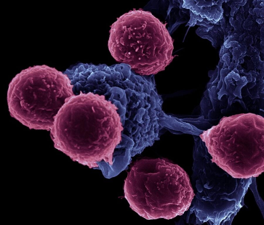
Our immune system acts as a shield to protect our bodies from foreign invasions of different kinds of pathogens. But what does the immune system contain? This is where we are going to discuss the cells of the immune system. The function of the immune cells is like soldiers protecting a country from the invading or foreign enemies. The discovery of immune cells started in the 19th century, the first of these was Elias Metchnikoff’s in 1883 discovered phagocytes and the process of phagocytosis. The discovery of the function of the thymus by Jacques Miller lead to the identification and function of T-lymphocytes and B-lymphocytes. These discoveries lead to a massive breakthrough in the study of immunologic cells and what roles they play.
Our immune system consists of an innate and adaptive system. The innate immune system is by birth whereas the adaptive system is acquired. The innate is also called non-specific immunity whereas the adaptive immune system is called specific immunity. Thus, the cells responsible for both specific as well as non-specific are known as leukocytes or the WBCs. All the leukocytes come from hematopoietic stem cells.
Hematopoietic stem cells are pluripotent stem cells that develop into all the blood cell types. They include both the WBCs and RBCs. They are found in the bone marrow. It was discovered in 1961 by Till and McCulloch in series of experiments which was conducted by them.
Let us look into the classification of immune cells in greater detail.
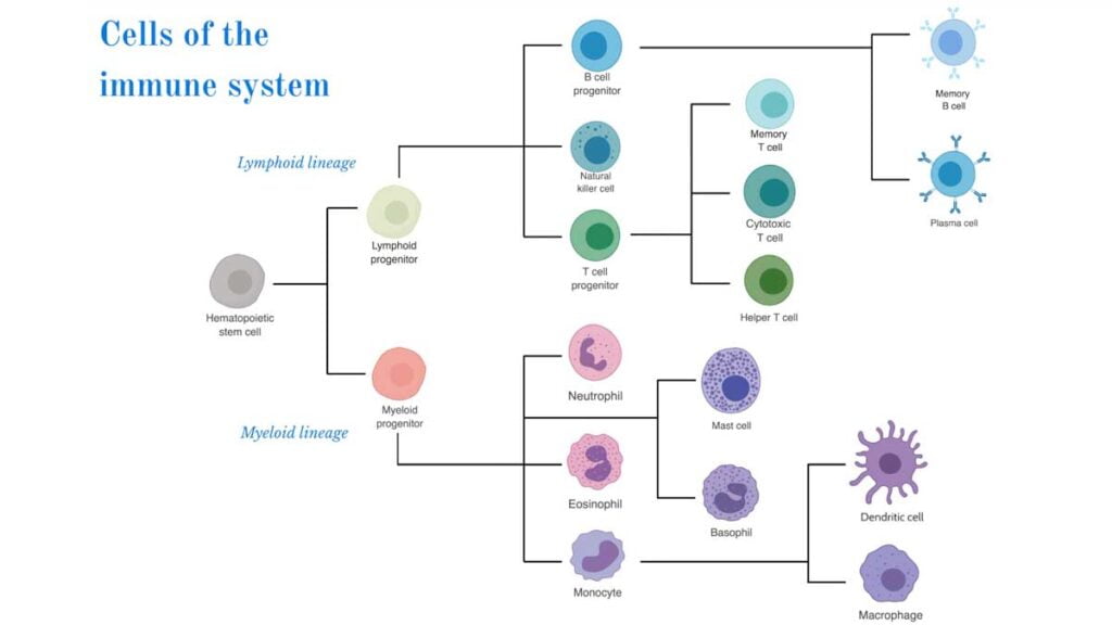
During hematopoiesis, the hematopoietic stem cells get differentiated along with one of the two pathways, giving rise to the common myeloid progenitor or the common lymphoid progenitor.
Common Lymphoid Progenitor
- The cells which arise from lymphoid progenitor are –
- B cell (B Lymphocytes)
- T cell (T Lymphocytes)
- NK cells
- These cells are responsible for the Adaptive immunity and are mononuclear leukocytes which comprise about 20-40% of total WBCs.
- They are found mostly in the blood, lymph and in lymphoid organs such as thymus, spleen, lymph nodes and appendix.
B Cells –
The B cells were discovered by Max Cooper and Robert Good in the 1960s in an experiment in which they demonstrated that the B cells were developed in the Bursa of Fabricius in birds. Jacques Miller found the presence of B-cells in thymus. In humans, the B cells are originated and matured in the bone marrow. The distribution is carried out in Spleen, lymph nodes, bone marrow, and other lymphoid tissue.
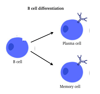
The naïve B cells also known as the immature B cells get proliferated and differentiated into cells which are –
1. Plasma cells (Effector B cells).
2. Memory cells.
The effector B cells carry out specific functions in order to eradicate and combat the pathogen. They are also known as plasma cells. They are short-lived and produce huge Antibodies against the pathogen. When an antigen is exposed, the production of effector cells (including T-cells) induces the primary response which then produces Antibodies against them. The antibodies cannot attack the pathogen or foreign substance which survives in the phagocytes.
The memory cells are long-lived cells that identify the foreign substance which had been previously exposed. When the memory cells identify the pathogen or foreign substance which had been previously encountered it gets differentiated into plasma cells and clear the pathogen. This induces the secondary response which is fast and instant.
The B cell mediated immune response against the microbes is also called humoral immunity.
T Cells –
- Jacques Miller had discovered the T lymphocytes in the Thymus.
- The T-cells arise in bone marrow but they do not get matured there, instead, they mature in the Thymus gland.
- The T-cell has a unique antigen-binding molecule called the T-cell receptors. These receptors are heterodimer in nature and consist of alpha and beta chains or gamma or delta chains.
- T-cells do not produce antibodies on their own but rather they perform various effector functions when Antigen-presenting cells bring the Antigen to the lymphoid organs.
- The T-cells generally help in eradicating the cancerous cells, self-altered cells, virus-infected cells, etc.
- The T-cells are generally divided into two major types –
The T cells are divided into two major types –
- T helper cells (TH cells)
- T cytotoxic cells (TC cells)
- Both of this are distinguished by the presence of the membrane glycoproteins in their cell receptors. The TH cells display CD4 whereas the TC cells display CD8 glycoproteins. The TCR (T cell receptors) present in the T cells do not recognize the free Antigens but instead it needs to bound to a particular class of self-molecule known as MHC (major Histocompatibility complex).
- The TH cells bound to MHC Class II molecules whereas the TC cells are bound to MHC class I.
- The effector and the memory T cell are generated after the activation of naïve T cells in the secondary lymphoid tissue.
- When naïve Tc cells are activated they differentiate into the cytotoxic T cells (CTL).
- When the naïve TH cells are activated they differentiate into variety of effector cells such as TH1, TH2, TH17, TREG etc. The differentiation subsets are due to distinct set of cytokines they produce.
- One of the main functions of helper T cells is to activate B cells to produce Antibodies and also Activate cytotoxic T cells by releasing cytokines. Thus, the cytotoxic T cells then kill the infected targets.
- The T-Cell mediated response is also called as cell-mediated response against the pathogen.
NK cells
- In 1975, Rolf Kiessling and his colleagues discovered that the killer cells which were genetically regulated, killed many types of tumors. Thus, named them as natural killer cells (NK cells).
- In general, NK cells develop in the bone marrow. They also develop in extracellular sites such as lymph nodes, Thymus, Spleen, etc.
- NK cells belong to the family of the innate lymphoid cell (ILC) which are large, granular, cytotoxic lymphocytes.
- These cells constitute about 5-10% of lymphocytes in human peripheral blood.
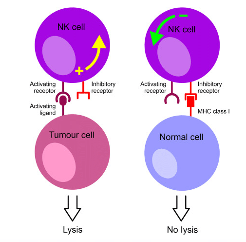
- They do not have membrane molecules and receptors but are still able to recognize the potential target cells. In some cases, an NK cell employs NK cell receptors to distinguish abnormalities, due to unusual reduction in the class I MHC molecule and unusual profile displayed by the receptors of the antigen of the infected cells by viruses or the tumor cells it is able to recognize. In another case, some infected cells or tumor cells have antibodies bound to their surface because the immune system has already produced an immune response, NK cells have a CD16 membrane receptor which helps to bind to those infected cells and this mechanism is also called antibody-dependent cell-mediated cytotoxicity (ADCC).
- One of the properties of NK cells is that they are capable to destroying malignant tumour and virus infected cells without any previous exposure or contact with the antigen.
- When there is absence of NK cells in the body, it is due to Chediak-Higashi syndrome which is an autosomal recessive disorder.
Common Myeloid Progenitor
This progenitor gives rise to numerous granulocytes like Basophiles, Neutrophils, Mast cells, Eosinophils. It also gives rise to monocytes such as macrophages and Dendritic cells and also the Erythroid progenitor. The Erythroid progenitor give rise to megakaryocytes and Erythrocytes.
1. Granulocytes –
- In 1846, Thomas Wharton jones represented “granule blood-cells” in several species including humans.
- They also are refered to as polymorphonuclear leukocytes, i.e. they have irregular shaped nuclei and are multilobed.
- They are generally WBCs classified as eosinophils, basophils, neutrophils, mast cells. The classification is based on their morphology and staining of their cytoplasmic granules.
a) Neutrophils –
- They were discovered by Elie Metchnikoff.
- They are also called as polymorphonuclear neutrophils.
- Constitute about 50-70% in the circulating WBCs.
- They are active phagocytic cells just like macrophages.
- They have multilobed nucleus and a granulated cytoplasm.
- As they contain granules, they release various enzymes which help to kill and digest the foreign cells.
- The identification of the characteristic cytoplasmic granules of neutrophils can be done by staining with both acidic and basic dyes.
b) Eosinophils –
- Discovered in 1874 by Heinrich Caro.
- The term ‘eosinophil’ was introduced by Ehrlich to describe cells with granules.
- They constitute about 2-5% of WBCs.
- They are motile and can move from blood to tissue space.
- They have a Bilobed nucleus.
- Their main role as an immune cell is defence against various protozoan and Helminth parasites.
- There are other roles such has killing foreign cells and controlling inflammatory responses.
- The identification of the characteristic of the cytoplasm of eosinophils can be done by using stain with acidic dye eosin.
c) Basophils –
- This type of cells was discovered by Paul Ehrlich in 1879.
- They constitute about not more than 1% of the total WBCs.
- They are non-phagocytic, were they release molecules which cause Allergic responses.
- Molecules which causes Allergic response, such as serotonin, Histamine, prostaglandins, etc.
- They posses high affinity receptors for antibody known as IgE.
d) Mast cells –
- Discovered by Paul Ehrlich in 1879.
- They are originated in the bone marrow.
- They get matured under the influence of C-kit ligand and stem cell factor in the presence of various growth factors.
- Just like Basophiles they also release molecules like Histamine.
- Other pharmacological active substance which they release are various cytokines, heparin, leukotrienes etc.
- They play a vital role in the development of Allergies.

2. Monocytes –
- Unlike Granulocytes, monocytes are mononuclear phagocytic leukocyte.
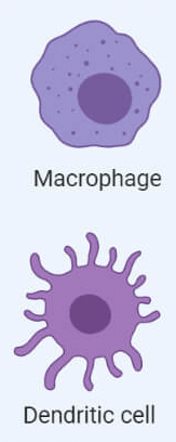
- They are produced in the bone marrow and further they circulate in the blood stream for about 8 hours and then migrate to tissue space and get differentiate into either macrophage or Dendritic cells.
- They are the largest type of leukocytes.
a) Macrophages –
- These cells were first discovered by Elias Metchnikoff in 1884.
- They are phagocytes and can be found in most of the tissues.
They have two origins –
- One from circulating monocytes which then migrate to tissue in response to the infection. They are known as inflammatory macrophages. Their function is have dual role in innate immunity: clear the pathogen directly and contribute their role and act as APCs (Antigen presenting cells).
- Other is from embryonic cells, known as tissue-resident macrophage like microglia in brain Kupffer cells in liver, Langerhans in the skin, Alveolar macrophage in the lung etc. adopt variety of tissue-specific functions.
b) Dendritic cells –
- They were discovered by Ralph Steinman and Zanvil Cohn in 1973.
- They Arise from both myeloid and lymphoid lineages.
- Their function is to capture Antigen and present them to other cells. They capture and acquire the Antigen by Phagocytosis, the Antigen the gets processed and then mature dendritic cells present them to TH cells.
- Dendritic cells express high level of class II MHC molecules.
They are generally classified into four types based on their origin and different function are:
- Langerhans cells
- Lymphoid dendritic cells
- Myeloid dendritic cells
- Interstitial dendritic cells
- Another type of Dendritic cell is the follicular dendritic cells which do not arise from bone marrow and have different function other than APCs. They do not express class II MHC molecule, thus do not act as APCs.
References –
Reference links –
https://www.ncbi.nlm.nih.gov/, , Kuby, J. and Kuby, J., 2007. Kuby immunology.
https://pathfinderacademy.in/book/csir-ugc-net-life-sciences-theory-practice-book-19.html


2 thoughts on “Immune Cells | Cells of Immune System”