Human Reproductive system follows Sexual mode of Reproduction where two genders are involved where their gametes fused to form offspring
Reproduction is a natural phenomenon and it is followed by living creatures on earth. Reproduction allows the living species to make like their own. And this allows to continue their existence on earth. The Reproduction can be sexual and asexual. To learn about Asexual reproduction click here. We human’s follow sexual type reproduction where two genders are involved and during fertilization, their gametes are fused to an embryo. In human reproduction, Male and Female are involved. Hence
Male Reproductive system –
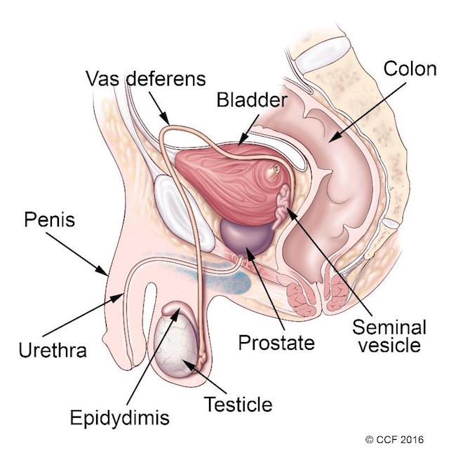
The male reproductive system consists of organs that are distributed inside (vas deferens, prostate and urethra) and outside the body (penis, scrotum and testis). The male reproductive system is connected to the urinary system via a common duct called urethra via which urine is passed and sperm is ejaculated. The urethra receives urine from the bladder and sperm from the testis. Similar to women, men’s reproduction system is monitored by sets of hormones released by the brain. The primary hormones are FSH and LH released by pituitary glands. FSH is required for initiating spermatogeneis and LH is responsible for the development of male characteristics.
The function of the male reproductive system is to release male sex hormones; synthesize and maintain the sperm and ejaculate it into the women’s reproductive tract.
The male reproductive system is made up of internal (inside your body) and external (outside your body) parts. Together, these organs help you urinate (rid your body of liquid waste materials), have sexual intercourse and make children
The male has a pair of testis and they are sites for spermatogenesis. The testes are oval-shaped organs present outside the body in the scrotum. The testis produces primary sex hormone i.e. testosterone which induces spermatogenesis and produce sperms. The testis works efficiently at 35℃ which is less than the body’s optimum temperature (37℃). This required temperature is maintained by the scrotum. The scrotum gets shrink during winter and swollen during summer to maintain the required temperature. The testicels have compartments and each compartment is called the testicular lobule. Each testicular lobule carries seminiferous tubules and this is where sperms are produced. The testicular lobule also carries a special type of cell called leydig cells which produces testosterone hormone. Once the sperms are ready, they travel via ducts called Rete testis. These rete testis are further opened in Vasa efferentia. The Vasa efferentia are further emptied in a highly coiled duct called Epididymis. It takes around 3 week to travel the sperm from seminiferous tubule to Epididymis during which it also get matured and motile.On sexual arousal, the sperm travel from testes via a duct called vas deferens. The vas deferens are associated with seminal vesicles (gland) and remain attached to the base of the bladder. The duct formed by the fusion of the vas deferens and seminal vesicle is called an ejaculatory duct that empties in the urethra. Another small walnut size gland called prostate gland is associated at the site of joining of the ejaculatory duct and urethra. Prostate gland function is to provide nutrition to the sperm for motility. Another pea shaped gland called Bulbourethral gland or cowper’s gland is associated with urethra just below the prostate gland. This gland produces lubricant for intercourse and acid neutraliser to reduce acidity of the urethra. The secretion released from seminal vesicles, prostate gland and bulbourethral gland is called accessory gland secretions. The accessory gland secretion and sperm are together called as semen.
Spermatogenesis –
Humans start making sperm when they reach puberty. The process is coordinated by hormones, which act as chemical messengers that ready the body for sexual maturity.
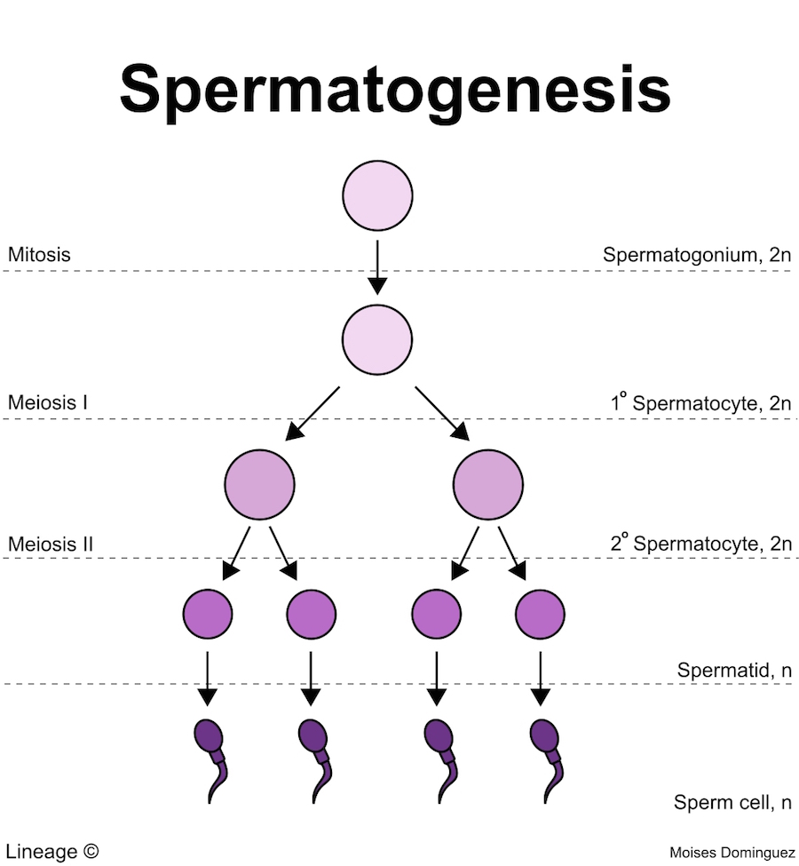
Spermatogenesis is a process of developing male gametes (sperm) from germ cells in the testes. This process is hormonally regulated and starts when the human men reaches puberty. When men reaches the puberty, hypothalamus glands releases Gonadotropin releasing hormones (GnRH). The GnRH stimulates anterior pituitary gland to release Follicle Stimulating Hormone (FSH) and Luteinizing Hormone (LH). Further, the LH causes Leydig cells to release testosterone and it further initiates spermatogenesis. And FSH stimulates sertoli cells to stimulate spermatogenesis and release nourishing elements to support sperm cells.
The spermatogonia (plural) or spermatogonium (Singular), the stem cells are present near the basement membrane of the seminiferous tubules. The seminiferous tubule also consists of leydig and sertoli cells. The sertoli cells provide nutrition to the growing sperms and leydig cells release testosterone. Some spermatogonia remain in an undifferentiated state and act as a reservoir for future sperm cells. The spermatogonium, a diploid (2n) cell undergo mitosis and produces and produces its replicate. Few spermatogonium loose the contact with the basement membrane of seminiferous tubule squeeze through tight junction of blood testis barrier. They further develop and differentiate into primary spermatocytes. The primary spermatocyte prepares itself for meiosis. The process starts with DNA replication and condenses the DNA. In meiosis I, the homologous pairs of chromosomes of the diploid cell align in the equator of the cell, followed by crossing over to exchange of genetic information. Meiotic spindle pulls the homologous pairs towards the pole and separates them. And it is followed by cell division forming two haploid secondary spermatocytes. Each spermatocyte carries 23 chromosomes but it has sister chromatids. These sister chromatids are separated in meiosis II forming four haploid cells called spermatids. These spermatids are further mature into spermatozoa or sperm. Hence, one primary spermatocyte produces four sperms. The whole process of formation of sperm from spermatogonium is called spermatogenesis.
Female Reproductive system-
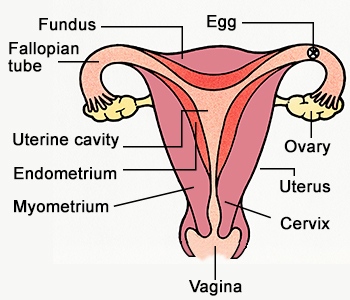
Compared to Men, women’s most of the reproductive parts inside the body. The female reproductive system consists of uterus, ovaries, fallopian tube, vagina and clitoris. The uterus is the site for the development of the baby. The uterus is mostly made of smooth muscle called myometrium which allows it to stretch during the baby’s development. Ovaries are female gonads, which produce ovarian hormones like estrogen and progesterone. And it is also the site for maturation of eggs.
Inside the uterus, there is an internal membrane is formed called endometrium. The endometrium has three layers namely stratum compactum, stratum spongiosum and stratum basalis. During the monthly menstrual cycle, every month, stratum functionalis is developed by stratum compactum and stratum spongiosum.
The female reproductive system has two ovaries and they site for oogenesis. They are small and almond shaped. They are induced by gonaotrophin released by the pituitary gland.The ovaries are made of connective tissue called tunica albuginea and squamous mesothelium. The cortex of the ovaries carries the ovarian follicles (immature egg).
Fallopian tube also called oviduct or uterine tube. It is the site of fertilization, where the egg awaits for sperm. The fallopian tube is attached to the ovaries via infundibulum, finger-like structure. The extended long thin wall structure is called ampulla and short with thicker wall structure is called isthmus. The intramural portion connects the fallopian tube with the uterine wall and opens it into the uterine lumen.
The vagina is a muscular tube-like structure which is an extension of the uterus and leads outside the body. Vagina is the site for sexual intercourse and it allows the penetration of penis and release of sperms. They are also made up of smooth muscles called stratified squamous, it is followed by elastic fibrous and smooth muscle making it stretchable. During intercourse, the vagina is lubricated with cervical mucus. The vagina and uterus are connected by cervix.
In the female body, urine is passed through the urethra and it is shorter than in men. It is beside the vagina. Female have an additional external genital called Clitoris is like reduced from of penis and its function is in sexual arousal.
Oogenesis-
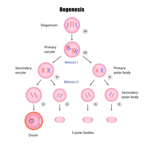
This process occurs in the female body to form haploid gametes (egg or ovum). The site for oogenesis is ovaries. The haploid gametes are formed from diploid germinal epithelium or primordial germ cell (they are kind of stem cells). The oogenesis process can be divided into three phases –
- Multiplication phase – In this phase, the germinal epithelial cells or primordial germ cell (2n) present in the ovary undergo mitosis and produce its million clones called gamete mother cells or oogonium cells. This phase occurs during the embryonic development of the female fetus inside the mother’s womb.
- Growth phase – In this phase, the mitosis stops and the gamete mother cells or oogonium starts growing in size and now it is called primary oocyte (2n). The primary oocytes initiate the meiosis I and get arrested in its prophase I. These arrested primary oocytes are stored in the ovaries.
- Maturation phase – This phase occurs at the puberty of the female body. And in this phase, the puberty hormones release the arrested primary oocytes and allows to complete the meiosis I process and forms two haploid daughter cells. But during this, their cytoplasmic division is unequal hence forming one larger cell called secondary oocyte (n) and another smaller called 1st polar body (n). The first polar body is non functional and degrades eventually. Now, the secondary oocyte continues the meiosis process and enters meiosis II. And again it gets arrested in metaphase II. This is when the ovulation occurs i.e. the arrested secondary oocyte at metaphase II undergoes ovulation. This arrest is released when the sperm comes into the contact. Further, the secondary oocyte completes its meiosis II and produces two daughter cells. Similar to meiosis I, one daughter cell is large called Ovum (mature oocyte) and other is small and it is called the 2nd polar body. The 2nd polar body eventually degraded. Hence, though one primary oocyte undergoes meiosis process and is expected to produce 4 gametes. But in reality it produces only one.
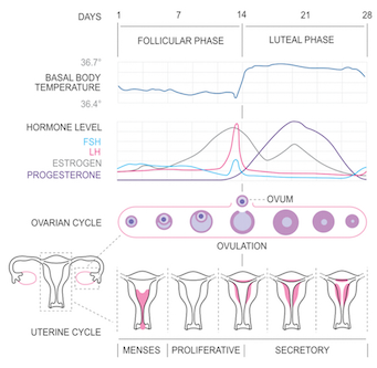
Ovarian and Menstrual Cycle in Female Reproductive System-
Ovorain and Menstrual cycle are both linked, interconnected cycles and occurs parallelly at the same time. These cycles are very vital for reproduction. Ovarian cycle occurs in ovaries and menstrual cycle happens in uterus. The cycle is monitored by the glands found in the brain namely Hypothalamus and pituitary gland.The hypothalamus releases Gonadotropin releasing Hormone (GnRH) which further stimulates pituitary gland. On stimulation, the pituitary gland releases Follicular stimulating hormone (FSH) and Luteinising hormone (LH). These hormones regulate the cycles. The levels of the hormones goes up and down throughout the cycle.
The ovary consists of primordial follicles carrying immature eggs. At birth, the girl child carries 400 000 primordial follicles in her ovaries. The follicles with immature eggs are called primary follicles. When the child reaches her puberty, these primary follicles on hormonal stimulation, provide a conducive environment to mature the egg. Every month, many follicles try to mature their egg but only one of them is able to mature its egg. The one who fails to mature their egg gets degraded. The follicle consists of granulosa cells which have receptors for FSH and LH. Hence, when hypothalamus stimulates the pituitary gland in response it releases FSH and LH which further stimulates granulosa Cells of follicles. This causes the proliferation of cells and growth of follicles. Once the follicle is mature it is called graffian follicle. During the maturation process, the follicle cells have the ability to produce estrogen hormone. Hence, more the number of follicle cells, higher is the concentration of estrogen in the blood. The high estrogen shows a positive feedback mechanism and induces pituitary gland to produce more LH. This is called LH surge which causes ovulation, where the egg cell is released from the follicle. The remains of follicle cells develop into corpus luteum. The corpus luteum has the ability to produce estrogen and progesterone. In return, the progesterone shows negative feedback on the pituitary gland and inhibit the production of FSH and LH.
The estrogen and progesterone monitor the menstrual cycle that occurs in the uterus. These hormones develop the thickening of endometrium lining and blood arteries. These hormones promote the proliferation of endometrium cells and blood arteries. The function of endometrium is to provide a nutrients and flourishing environment for the fertilised egg and get implanted inorder to further develop into a fetus.
In case, when there is absence of sperm, there is no fertilization. The corpus luteum starts degrading and reduces the amount of progesterone and estrogen causing degeneration of endometrium; this gives menses or bleeing via vagina. The low concentration of progesterone and estrogen will also induce the production of FSH and LH. The high FSH and LH further induces primary follicle maturation to secondary follicle.
Fertilization in Human Reproductive System-
It is defined as the fusion of the male and female gametes. This process happens only after the sexual intercourse where the sperms in the form of semen are ejacuated in the female reproductive tract. When sperms enter via vagina, it travels from vagina to cervix, cervix to uterus and uterus to fallopian tube. In the fallopian tube, the egg waits for the sperm and there they get fused together.
The semen is composed of millions of sperms, water, fructose, citrate, fibrinogen, mucus, prostaglandins and buffers. The water acts as a medium in which sperms are suspended. Fructose and citrate acts like an energy source. Fibrinogen helps the sperm to get attached to the wall of the uterine lining. Mucus acts as a lubricant and buffers to neutralise pH.
As soon as the semen is introduced into the vagina, the sperm starts moving towards the uterus. The prostaglandins cause the uterus contraction which pushes the sperm inside the uterus. An additional female hormone called oxytocin released by her pituitary gland is also responsible for contraction of the uterus wall. While travelling towards the fallopian tube, many of the sperm die off and only few thousands of sperms are able to reach the fallopian tube. These sperms undergo a process called capacitation. The sperm that reaches the egg before capacitation cannot fertilize the egg. If a female is ovulated, the egg waits in the fallopian tube for fusion with sperm. The capacitation process occurs only inside the female body. The uterus has chemical substances like albumin, lipoproteins, proteolytic and enzymes and they are involved in capacitation. The capacitation process is characterised by the changes in structure of the sperm like protein phosphorylation, enzyme activity, activation of metabolism and membrane structure making it hyperactive and motile. The capacitation process also exposes the sperm’s ligand which will further bind to the egg’s receptor.
Once the sperm reaches the egg, the sperm releases its hydrolytic enzymes like hyluronidase. This enzyme will break the cumulus matrix (follicle cells and Hyluronic acid) of the egg cell and try to penetrate it. The egg has a receptor on the Zona pellucida surface for the attachment of the sperm. The penetration allows the sperm to reach the receptor and bind it. The ligand-receptor binding stimulates the fusion of Sperm plasma membrane with its acrosomal membrane releasing the hydrolytic enzymes. These enzymes will degrade zona pellucida and expose the egg’s plasma membrane. Due to the release of acrosomal enzymes, the acrosomal membrane of sperms gets exposed. The acrosomal membrane has ligands which are specific for the receptor present on the egg’s plasma membrane. This recruits the smooth endoplasmic reticulum to release the calcium ions causing the polarization of the egg’s plasma membrane making the membrane positive at the inner side and negative outside.
The polarisation of membrane inhibits and blocks the binding and penetration of other sperm into the egg cell. The other mechanism of blocking other sperm is breaking the cortical granules which release some enzymes that break the zona pellucida and its receptors. Hence, no other sperm can bind it. During this, the sperm that has successfully penetrated the egg cell, releases its nucleus in the egg cell.
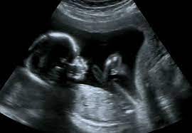
The released calcium ions also release the egg cell from metaphase II arrest forming one mature ovum and 2nd polar body. The mature ovum then fuses with the nucleus of the sperm cell forming diploid zygote. The zygote will further develop into an embryo and then to a fetus. Whereas, the polar body will be degraded.
Blastulation and Gastrulation stages of Fertilization of Human Reproductive system-
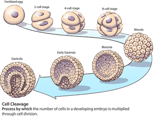
This process starts when the sperm cell nucleus fuses with mature ovum forming a diploid zygote. The zygote is a totipotent stem cell which has the ability to form a baby. The zygote further undergoes mitosis and increases the cell number from 1 to 2, 2 to 4 and 4 to 8. The zygote with 4 cells and 8 cells are called the 4 cell stage and 8 cell stage respectively. The 8 cells stage further forms 16 cells and this stage is called Morulla. The stages from 2 cells to 16 cells are altogether called cleavage steps. The cells inside the Morulla are called Blastomeres. The Morulla stage is further developed into Blastocyst, where the blastomeres get rearranged. Few blastomeres arrange themselves at the boundary and now they are called outer cell mass. While few get assembled at one side forming inner cell mass. The empty cavity is called blastocoel. The outer cell mass and inner cell mass are further developed into trophoblast and embryoblast respectively. The embryoblast develops into embryo and trophoblast forms the placenta. The function of placenta is to provide nutrients to the developing baby. This stage of the development is called blastocyst. The process of embryo development till the blastocyst stage is called Blastulation. The blastulation process goes for 2 weeks.
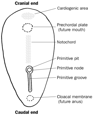
Once the blastocyst is formed, it travels from the fallopian tube to the uterus. The trophoblast cells help the blastocyst to get attached to the endometrium of the uterus. The attachment of blastocyst to the endometrium is called implantation. This is when the trophoblast forms placenta to provide nutrients to the growing embryo. The inner cell mass starts arranging first into two layers and also divides the blastocoel cavity into two halves. The upper layer of inner cell mass layer is called epiblast and the lower is called Hypoblast. The cavity about two cell layers is called Amniotic cavity and the cavity below the cell layer is called Yolk sac. The circular shape oval has two poles, one is called cranial end and other is called caudal end.
In the Epiblast layer, some cells start arranging into a streak and node called primitive streak and primitive pit respectively. Though the streak appears at the cranial end first but later it starts developing towards the caudal end forming a primitive groove; a primitive node develops into a primitive pit. The development of the streak distinguishes cranial and caudal ends. The cranial ends develop the head and mouth and caudal ends develop the anus.. The cells of the primitive grove start multiplying and form an additional third layer called mesoderm. This process is very complex. And during this the epiblast starts replacing hypoblast and forms the endoderm layer. And at this stage, the epiderm is called endoderm. The formation of three primary layer of germ cell structure is called Gastrulation. The primitive pit cells also start multiplying and form notochord in the mesoderm layer. During the baby’s development, the notochord forms the nervous system.
In every week, different organs are developed inside the mother’s womb and It takes 9 months to develop the baby.
References –
https://www.histology.leeds.ac.uk/female/FRS_ovarian_fol.php
Tortora, G. and Derrickson, B., 2012. Introduction to the human body. 9th ed. Hoboken, NJ: John Wiley & Sons.
Dr. Sangha Bijekar has 9 years of Teaching Experience at University level. She loves to get engage in teaching and learning process. She is into blogging from last two years. She intends to provide student friendly reading material. She is avid Dog Lover and animal rescuer. She is learned Bharatnatyam and Katthak Dancer. She is into biking and She also loves to cook.