Composition of bacterial cell wall is crucial to understand as it gives shape, rigidity, and support to the cell.
Introduction to Composition of Bacterial Cell Wall
Bacteria are prokaryotic cell. They are unicellular. They lack membrane bound organelle and Nucleus. The Genetic material is present in the form of nucleoid. The cell’s internal structure and components are protected by the rigid cell wall.
It has found that almost 90% bacteria have cell wall but few lack it. The function of the rigid cell wall is to protect the cell from an external osmotic pressure.
As per the osmosis principle, the ions, solute or molecules always move from higher to lower concentration. Compared to outside environment, the cell has high solute concentration and the outside environment is diluted. And hence as per the osmosis principle, the solute from cell would move outside and water from outside environment would enter the cell. This may cause cell lysis. The cell wall protects the cell from osmotic lysis.
The study has also found that certain pathogenic bacterial cell wall is responsible for pathogenicity (disease causing ability). The difference in the cell wall composition is also used for identification and classification of bacteria. Based on the composition of cell wall, the bacteria are broadly classified as Gram positive and Gram Negative.
Structure of Bacterial Cell wall:
- In bacteria, the cell wall is made from peptidoglycan.
- It is also called as murien. It is a mesh like structure made from alternating residues N-acetyl glucosamine (NAG) and N-acetylmuramic acid (NAM) linked by of β-(1,4) glycosidic bond.
- NAG and NAM are an amide derivative form of carbohydrate. A short chain of 3 to 5 amino acids are linked to each N-acetylmuramic acid (NAM) of NAG-NAM polymer.
- The type and number of amino acid may differ from bacteria to bacteria. The amino acids are linked to each other by peptide bond.
- The short chain hanged on each N-acetylmuramic acid (NAM) is also crosslinked to each other by pentaglycine (5 glycine).
Such mesh like structure makes the cell wall rigid and strong.
Difference between Gram positive and Gram Negative
History of Gram Staining:
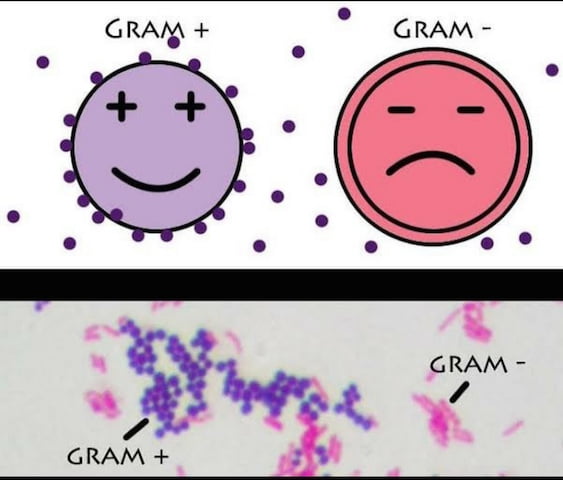
In 1882, Hans Christian gram invented the staining technique for visualization of pneumonia causing agents. His technique was inspired from Robert Koch staining method. Christian Gram modified the staining technique by using crystal violet as a primary stain, iodine as a mordant and alcohol as decolorizing agent. While staining the lung cells, Christian Gram found that the host cells remained unstained. And the pathogenic bacteria appeared circular in shape and blue or violet in color. And hence, the bacteria taking Gram stain were called as Gram-positive.
Christian Gram continued to use this staining technique to stain and visualize series of bacteria. While conducting staining experiments, Gram found that Typhoid causing bacteria did not appear as blue or crystal violet. He found and understood that the crystal violet color is decolorizing the primary stain. And hence, the bacteria that did not take Gram stain were called as Gram-negative bacteria.
This technique is still widely used for staining and distinguishing the bacteria. It is one of the primary staining method that is used in the filed of Microbiology. Let’s see the difference between the cell wall composition and structure of Gram-positive and Gram-negative bacteria. This difference is the cause for taking two different stains.
Cell Wall of Gram Positive Bacteria
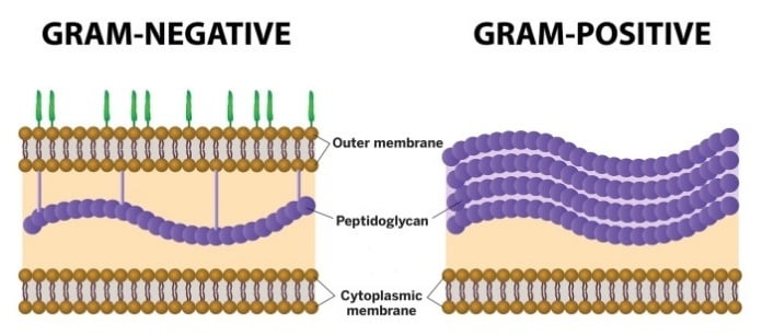
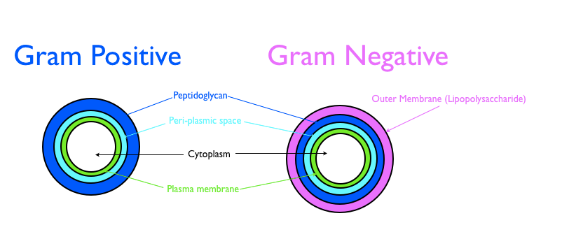
- In gram-positive bacteria, the peptidoglycan layer is thick.
- It contains teichoic acid chains that are extended from cell membrane to peptidoglycan.
- Teichoic acids are bacterial polusacharides derivates.
- Teichoic acid are copolymers of glycerol phosphate or ribitol phosphate linked via phosphodiester bonds.
- Due to the phosphate group, it contains negative charge and hence it attracts positive ions like magnesium and sodium.
- The presence of teichoic acids provides additional rigidity to cell wall.
- Research has also shown that presence of teichoic acids leads to increase in pathogenecity of bacteria.
- Some gram positive have additional coating of mycolic acid.
- The prominent example is Mycobacterium tuberculosis. This bacterium is very well known for causing tuberculosis. Such gram-positive bacteria need to be stain by special method called as acid fast staining.
One of the mechanisms of causing disease of pathogenic gram positive bacteria is synthesizing and releasing toxins in the host body. As these toxins are released out it is called as exotoxins.
Cell Wall of Gram negative Bacteria
- The Gram-negative bacterial cell wall is more complex than gram positive bacteria.
- The Gram negative bacteria also contains peptidoglycans but it is thin as compared to Gram-positive bacteria.
- In gram Negative bacteria, the peptidoglycan layer is externally covered with outer membrane.
- This outer membrane is made from lipopolysacharides (LPS).
- The LPS is made from three components – Lipid A, a core polysaccharide, and an O antigen.
- The presence of LPS provides additional protection to the bacterial cell.
- The hydrophobic Lipid A is anchored in cell membrane of bacteria.
- The core polysaccharide is attached to lipd A.
- The other end of core polysaccharide is liked to O Antigen (glycan polymer/carbohydrate).
- The presence of O antigen cause smooth appearance of Lipopolysaccharides (LPS).
- Gram-negative bacteria with O antigen (smooth LPS) are more resistant to antibiotics than rough LPS (absence of O Antigen). The absence of O antigen makes it permeable to hydrophobic antibiotics.
- The composition of core polysacharride and O antigen may vary from bacterial strain to strain and hence this data can be used for bacterial classification.
- The outer membrance and plasma membrane are separated by periplasmic space.
- When the host immune system attack and lysed the gram negative bacteria.
- The lysis of bacteria causes release of lipid A in the blood stream. The lipid A acts like endotoxin and may leads to endotoxin shock or septic shock to host.

Meme –

Activity for understanding the Composition of Bacterial Cell Wall:
Distinguished between Gram positive and Gram negative bacteria based on following characteristics-
| Cell wall Composition | Characteristics | Gram Positive Bacteria | Gram Negative Bacteria |
| Peptidoglycan | Thick or Thin? | ||
| Rigidity of cell wall | Which bacterial cell wall is more rigid? | ||
| Teichoic acid | Present or Absent | ||
| Outer membrane | Present or Absent | ||
| Mycolic acid | Present or Absent | ||
| Periplasmic space | Present or Absent | ||
| Toxins | Endo to Exo type? |
Acknowledgement –
The Biokimicroki team really appreciate Amrita Dinesh for contributing the meme for Composition of Bacterial Cell Wall article.
References –
https://bio.libretexts.org/Bookshelves/Microbiology/
https://asm.org/getattachment/5c95a063-326b-4b2f-98ce-001de9a5ece3/gram-stain-protocol-2886.pdf
http://www.textbookofbacteriology.net/structure_5.html
Dr. Sangha Bijekar has 9 years of Teaching Experience at University level. She loves to get engage in teaching and learning process. She is into blogging from last two years. She intends to provide student friendly reading material. She is avid Dog Lover and animal rescuer. She is learned Bharatnatyam and Katthak Dancer. She is into biking and She also loves to cook.


