“Plasmolysis term is derived from latin word plasma meaning matrix and lysis means loosening i.e. loosening of cell matrix”.
Aim –
To study and observe the plasmolysis phenomena of plant cells in the laboratory conditions.
Theory –
What is Plasmolysis?
Plasmolysis is the process that describes the loss of water resulting in shrinkage of the cell. This happens when the cell is place hypertonic solution. The hypertonic solution is a solution that contains higher concentration of solute than the suspended cell. Plasmolysis occurs due the semi-permeable nature of cytoplasmic membrane. In plasmolysis, the cell and its cytoplasmic content gets shrink. When the cell is placed in hypertonic solution, the water present inside the cell moves out causing cell shrinkage. And when the cell is placed in hypotonic solution, the water from solution moves inside the cell causing swelling of the cell. Extreme swelling and shrinkage of cell may lead to the cell death.
Plasmolysis and Osmosis –
The principle behind the plasmolysis is the osmosis. Osmosis is the movement of water across the semi permeable membrane from higher concentration to lower concentration. The plasmolysis does not occur in natural conditions but can be observed in laboratory conditions. The state or condition when cell undergo plasmolysis it is called plasmolyzed cell.
How water permeates via cell membrane?
The cytoplasmic membrane is semi-permeable in nature. The semi permeable membrane selectively allows the entry and exit of water and essential ions and inhibiting other. Its function is to maintain cell turgidity. The turgidity is defined as the swollen state of plant cell. The turgidity of plant cell is responsible for keeping the plant stand upright without any skeletal support. The water present inside the cell generate turgor pressure against the cell wall. In natural condition, such turgid cell prevent the further intake of water.
Principle –
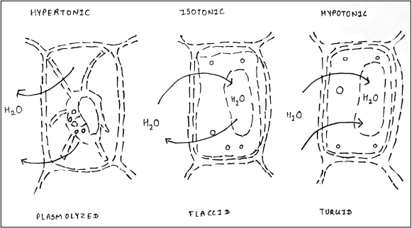
When the plant cell/s is/are suspended in solution of different solute concentration, their behaviour varies. The Hypertonic solution is a solution that contains higher concentration of solute than found inside the cell. Isotonic solution contains equal concentration of solute to cell. Hypotonic solution contains lesser concentration of solute than cell. In a hypertonic solution, the cell contains more water than the solution and therefore the water
molecules move out and cause the cell to shrink. In an isotonic solution, as there is an equilibrium of solute in and outside the cell, there is no change in the cell shape.
Stages of Plasmolysis –
- Incipient Plasmolysis – It is the first stage, where there is not much change in the cellular morphology. Due to hypertonic solution, the water from cytoplasm starts flowing out from the cell. Due to which, the cytoplasm shrink and protoplast moves away or get detached from the cell wall. This stage is reversible and become flaccid.
- Evident Plasmolysis – This is the second stage, where the protoplast gets shrink and detached from cell wall to its maximum extent. This stage is also reversible.
- Final Plasmolysis – This is the last and final stage where the protoplast get detached from cell wall completely and moves to the center of the cell.
Types of plasmolysis based on its appearance –
- Conclave plasmolysis – as the name suggest that water efflux from the cytoplasm results in the formation of conclave shape protoplast. It is formed due to the partial detachment of cytoplasmic membrane from the cell wall. it appears like half moon shape.
- Convex plasmolysis – When there is excessive water loss, the protoplast shrink and get completely detached from the cell wall resulting in convex shape protoplast inside the cell. The convex plasmolysis is irreversible and lead to cell death.
Requirements –
- Plant sample (Rhoeo leaves) – because these colorful leaves allows to observe plasmolysis clearly.
- Forceps
- Needle
- Watch glass
- Droppers
- Glass slides
- Cover slip
- Compound microscope
- Sodium chloride (0.1% and 0.5%)
Procedure –
- Wash, clean and dry the glass slide and place it on your working bench.
- Select fresh leaves, wash them and place them on watch glass.
- Fold the leaves in order to tear them lower side with the help of blade.
- Using forceps remove the transparent layer. The purpose of tearing and removing layer is to expose the epidermis layer of the leaf.
- Take the epidermal layer and place of it on glass slide.
- Using dropper, add 1 or 2 drops of 0.1% sodium chlorides solution on the placed epidermal layer of glass slide.
- Further again add
- 1 or 2 drops of 0.5% sodium chlorides solution on the same slide.
- Now carefully cover the sample with the coverslip. No bubble should be formed.
- Keep the slide undisturbed for few minutes.
- Observe the slide under microscope.
You can watch the protocol here –
Observation –
When we observe the slide under microscope, the plant cells become flaccid in 0.1% of sodium chloride and become shrink in 5% of sodium chloride.
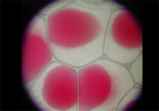
Conclusion –
The performed experiment shows that the plant cells exhibit plasmolysis phenomenon. When the cell is immersed in the hypertonic solution (0.1% and 5% of sodium chloride), the cell loses its water resulting in the shrinkage of the cytoplasmic content.
Dr. Sangha Bijekar has 9 years of Teaching Experience at University level. She loves to get engage in teaching and learning process. She is into blogging from last two years. She intends to provide student friendly reading material. She is avid Dog Lover and animal rescuer. She is learned Bharatnatyam and Katthak Dancer. She is into biking and She also loves to cook.