Yeasts are unicellular prokaryotic organisms. They cannot be seen by naked eyes and hence we need to observe them microscopically.
Introduction:
Yeasts are unicellular and microscopic fungi. They are commercially used in bread and wine making industries. They multiply asexually by following budding method. The shape and size varies from species to species of yeast and they are found in the form of spherical, elongated or egg shaped type. The yeast are non-motile organism because they lack flagella or any other locomotary organelle. As it is an eukaryote, it contains mitochondria, Endoplasmic reticulum and nucleus in the cytoplasm.
A temporary slide is prepared by placing the desired cell suspension on the slide. The cell suspension can be either stained or directly observed. The cell suspension is covered by cover slip with no air bubble trapped. The slide with microbial cell suspension covered with cover slip is observed under microscope.
Principle:
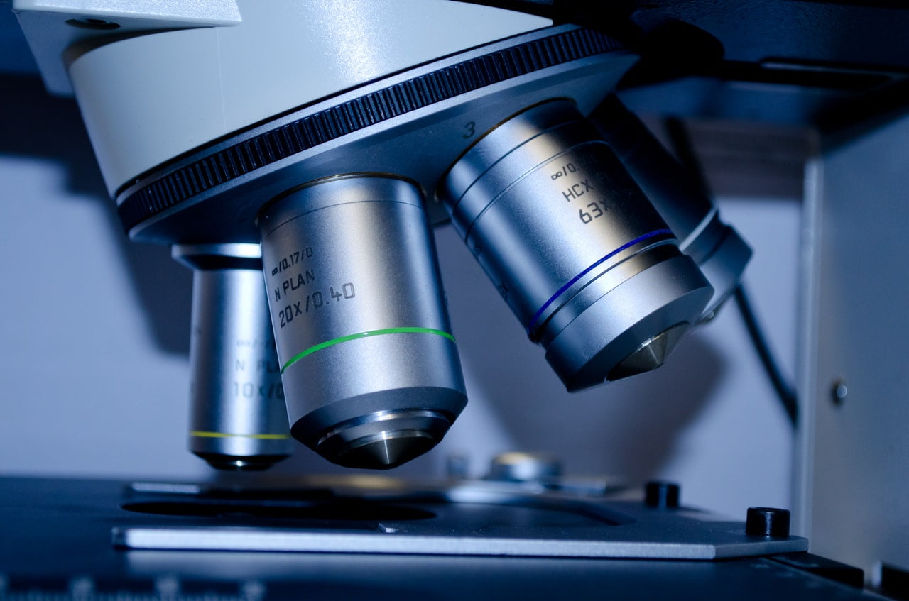
The microscope slides are thin, flat and transparent and made from glass. The microbial sample is placed on the slide and covered with cover slip and then observed under microscope. Their suspension is mounted on slide and focused to observe its morphology.
Requirements:
- Slide
- Cover slip
- Dry yeasts
- Sugar
- Conical flask
- Water
- Bunsen burner
- Inoculating loop
Procedure:
- Boil 50 ml of water in conical flask and allow it cool warm it down.
- Add spoonful of sugar and dissolve it
- Add dry yeasts in warm water and mix properly.
- Cover the conical flask for 15 min.
- In 15 minutes, the yeast gets activated.
- With the help of sterile inoculating loop, a loopful of yeast is mount on slide.
- Cover slip is placed on top and observed under microscope by using different modification.
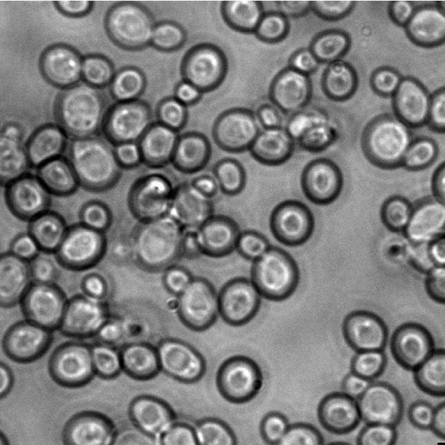
Observation of Yeast:
Active yeast cells and protozoa cells were observed under microscope
Result:
Yeasts are unicellular eukaryotic organisms and their morphological studies can be done by light microscope.
References –