This article explains the principle and procedure of Gram staining technique. The principle and procedure of gram staining is based on the difference in the cell wall composition leads to adsorption of different stains. In Gram staining procedure, two different stains are used. Gram staining technique is used to differentiate the bacteria. The gram Staining method was invented by Hans Christian Gram.
Introduction of Gram Staining Technique
In 1882, Hans Christian gram invented the staining technique and published in 1884. In1921, this technique was further developed by Hucker. This staining was named as gram staining to honor the contribution of Hans Christian Gram. The Gram staining technique is used for visualization and differentiating bacteria based on the composition of cell wall.
Read our article on Composition and Difference in Bacterial Cell Wall for better understanding of Gram Staining.
Principle of Gram Staining Technique:
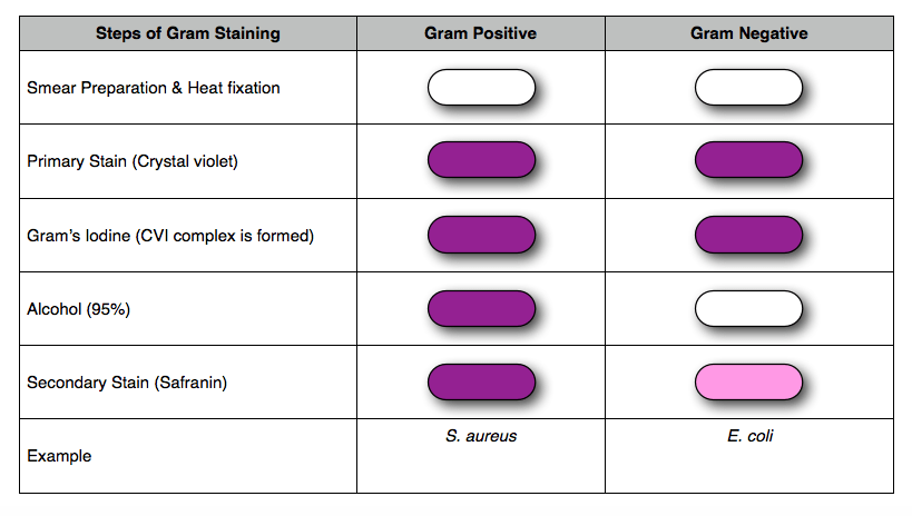
The Gram-negative bacterial cell wall is more complex than Gram-positive bacteria. The bacterial cell wall is composed of peptidoglycan. The Gram-positive bacteria have thick peptidoglycan while Gram-negative bacteria have thin peptidoglycan layer. When crystal violet, the primary stain is flooded on bacterial cells, the cell wall takes the primary stain and both the Gram positive and Gram-negative bacterial cells appear as violet or blue color.
The mordant (iodine) forms a complex with crystal violet stain present in the cell wall and forms crystal violet Iodine complex (CVI complex). When the bacterial cells are decolorized with 95% alcohol, the lipid part of lipopolysaccharide (outer membrane) of Gram-negative cells gets washed away. The lipid gets washed away because they are soluble in alcohol. With these lipids, the crystal violet also gets washed off in Gram- negative cells and making Gram-negative cells almost colorless.
When secondary stain, Safranin is flooded, the colorless Gram-negative cells take the Safranin stain and appear pink in color. As the Gram-positive bacteria have already adsorbed the crystal violet color in their cell wall, they cannot take any secondary stain. And hence, they appear blue or violet in color.
Requirements for Gram Staining
- Bacterial culture
- Slide
- Cover slip
- Dropper
- Distill water
- Inoculating look
- Burner
- Primary stain – Gram’s Crystal violet
- Secondary stain – Gram’s Safranin
- Gram’s Iodine
- Alcohol
Procedure:
- Wash and clean glass slide (make it dust and grease free)
- Dry the glass slide.
- With the help of inoculating loop, take a loopful of culture and spread it uniformly on the slide, this step is called as smear preparation.
- Allow the smear to dry in air and then heat fixed it. Heat fixing kills bacteria and adheres them to the slide.
- Flood the crystal violet (primary stain) on the smear for a minute.
- Run off the crystal violet by using distilled water.
- Gently flood gram’s iodine over smear for a minute. At this moment the smear would appear violet or blue in colour.
- Decolourise the smear by flooding the slide with 95% alcohol for 30 sec.
- Flood the safranin (counter stain) over the slide and keep it for a minute.
- Run off the safranin stain with distilled water.
- Allow the slide to dry and observe under microscope.

Observation:
Either pink or violet color Bacteria would be observed under microscope
Result:
Pink color Bacteria indicates that the given bacteria are gram negative and violet color indicates that the given bacteria are gram positive.
Conclusion:
Gram staining technique differentiates bacteria into two large groups on the basis of composition of cell wall. Hence, it is one of the primary methods used for identification of bacteria.
References:
https://serc.carleton.edu/microbelife/research_methods/microscopy/gramstain.html
https://www.diffen.com/difference/Gram-negative_Bacteria_vs_Gram-positive_Bacteria
http://vlab.amrita.edu/?sub=3&brch=73&sim=208&cnt=2


3 thoughts on “Principle and Procedure of Gram Staining Technique”